Discography
See clearly so you can treat optimally
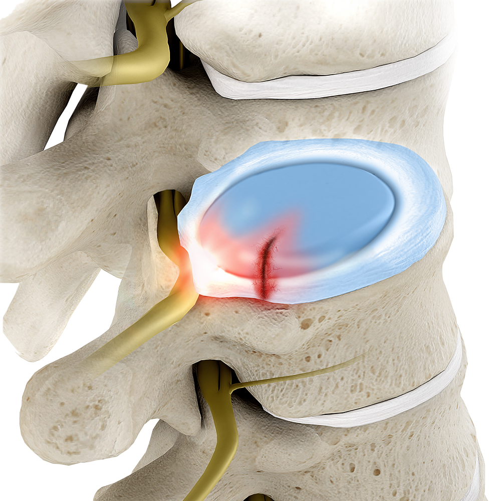
When patients are in pain, the first thing they need is answers. That’s what discography helps you provide. By injecting dye into one or more discs while monitoring for reproduction of back pain, this imaging procedure empowers you to identify exactly which of the discs is the source of pain. Beyond identification, discography can detect structural damage in a disc and reveal whether a disc has begun to rupture or has tears in its outer ring—assisting with treatment planning.1
Benefits
Diagnostic accuracy studies show strong evidence that discography is a useful tool for imaging disc morphology missed by MRI and other tests.3
How it works
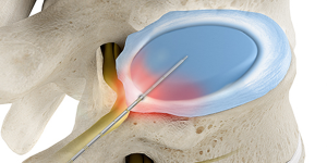
Under x-ray guidance, a thin guide needle is inserted into one or more discs. The discs are then pressurized one at a time with contrast dye to outline any damaged areas. If your patient experiences pain that feels like their usual pain, that disc can be classified as a pain-generating or symptomatic disc. By asking questions about the intensity, type, and location of the pressure, you can determine your diagnosis and appropriate treatment options.1
Procedure14
1
Discography typically takes about 30-60 minutes to perform. During that time, your patient will be awake but sedated. This allows them to communicate what they are feeling as the procedure progresses.
2
A local anesthetic is used to numb the skin and all the tissue down to the disc area. Using x-ray guidance, a needle is inserted into the center of the disc. This process may be repeated for multiple discs. Once all the needles are placed, the discs are pressurized one at a time with injections of contrast dye. With each injection, the patient will feel either pressure or pain. If they feel pain, you can ask how it compares to the usual pain.
3
After each disc is tested, x-rays are taken and the needles are removed. The patient may be taken for a CT scan to obtain additional information about the exact pattern spread of dye through or out of the disc.